English guidance
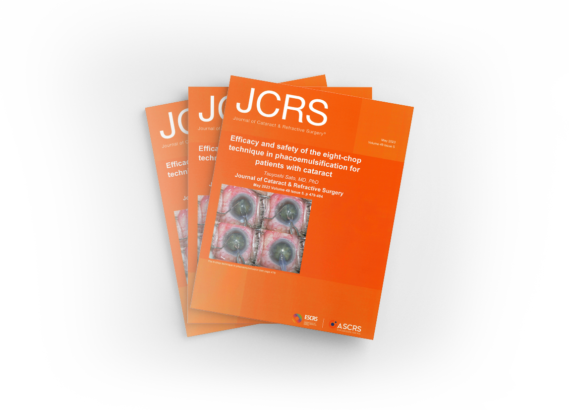
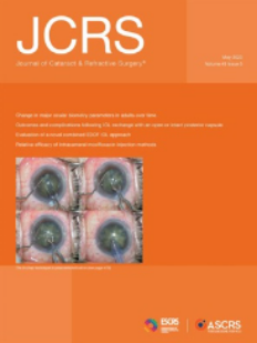
The eight-chop technique was featured on the cover of the Journal of Cataract & Refractive Surgery (JCRS).
The JCRS journal is published by the American Society of Cataract and Refractive Surgery and the European Cataract and Refractive Society.
As of 2023, the eight-chop technique was listed as the most up-to-date cataract technique in the magazine, the eight-chop procedure graced the cover of the magazine.
Also as of July 2023, most high-profile article in the JCRS journal.
"Eight-chop technique" and "Lance-chop technique"
EIGHT-CHOP TECHNIQUE
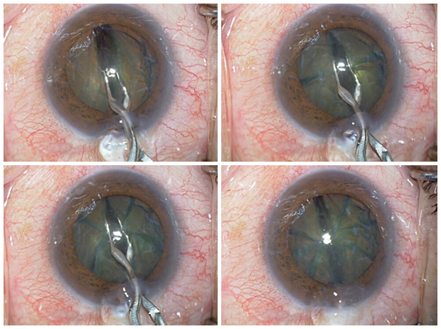
Safety-oriented advanced surgery
In 2002 This technique was developed by our Director, Tsuyoshi Sato.
It is a development of the prechop technique, which is technically the most difficult technique in cataract surgery, and involves dividing the lens nucleus into up to eight sections.
It was presented at the Ophthalmic Surgery Congress in 2009 and named the eight-chop technique.
In 2019, we also developed the Lance Chopper and completed the lance-chop technique, which was presented at the Japanese Society for Ophthalmic Surgery.
LANCE-CHOP TECHNIQUE
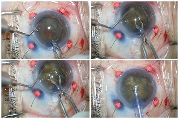
Safe surgery in difficult cases
It is used when the lens is hard and cannot be divided by the eight-chop technique.
- It may also be used when the lens nucleus is not hard but the division of the lens nucleus is incomplete.
- This technique is essential in refractory cases such as weak zonule, corneal opacity and small pupill.
- It can be safely performed in most cases where other techniques are not possible.
DEVELOPMENTS OF EIGHT-CHOPPER
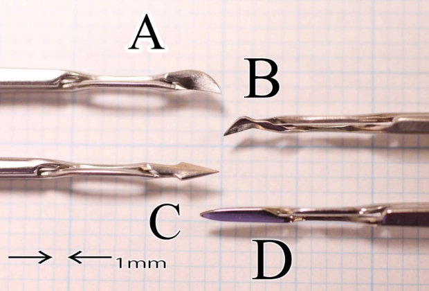
B: Eight-chopper II
(ASICO:SP-8402)
C: Lance-chopper
(ASICO:SP-9989)
Sustainer(ASICO:AE-2530)
-
Eight-chopper I
The prechopper, which divides the lens nucleus into four sections, was unsuitable for operation in narrow sacs due to its large tip when performing multiple sections, so the length of the tip was reduced (3.2 mm, width 1.4 mm) and the leading edge was made sharper.
The operability has been improved and the lens nucleus can now be divided without difficulty.
The effectiveness of surgery with the eight-chopper I was presented at the Ophthalmic Surgery Society in 2009. -
Eight-chopper II
The Eight-chopper II was developed by downsizing and sharpening the tip of the Eight-chopper I. The tip is 2.5 mm long and 0.8 mm wide, with a sharp leading edge. The tip is angled so that it can be inserted vertically into the lens nucleus. With the Eight-chopper II, the eight-chop technique can be used for harder lens nuclei in minimal incision cataract surgery with a corneal incision wound of 2.3 mm.
The usefulness of the Eight-chopper II in minimal incision cataract surgery was presented at the Japanese Cataract Society in 2011. -
Lance-chopper
Used when the lens nucleus is very hard. Conventional universal prechoppers were difficult to insert and the long tip made operation difficult.
The tip is 3.0 mm long and 1.3 mm wide, with a sharp leading edge. The short and sharp tip facilitates intraocular manipulation, makes it easier to penetrate the lens nucleus and the wide tip enables the fissure to be enlarged by pressing on the cross-section of the lens nucleus.
The effectiveness of surgery with the Lance-chopper was presented at the Ophthalmic Surgery Society in 2019.
DOCTOR REFERRAL

TSUYOSHI SATO
- Member of Japanese Ophthalmological Society
- Member of Japanese Society of Ophthalmic Surgery
- Member of Japanese Society of Cataract and Refractive Surgery
- Member of American Society of Cataract and Refractive Surgery
- Member of European Society of Cataract and Refractive Surgery
Director Biography
The degree of Doctor of Medicine was awarded by Dr. Leo Esaki, President of the University of Tsukuba (Nobel Prize winner), after submitting a dissertation on the retinal circulation to the University of Tsukuba. During my study abroad period, I was also taught by Dr. Sayon Roy, Professor of Boston University School of Medicine, a world authority on diabetic retinopathy, and my paper was published in Diabetes, the premier medical journal in diabetes research. In addition, I was the first in the world to identify one of the pathogenesis of glaucoma caused by diabetes, and my paper was published in Investigative Ophtalmology & Visual Science, the premier medical journal in ophthalmology research.
History
Graduated from Tsukuba University
Obtained a medical license
Obtained a medical degree in ophthalmology
Received a medical degree (Doctor of Medicine)
Career History
Resident,Department of Ophthalmology, University of Tsukuba Hospital
Clinical and Research Instructor, Department of Ophthalmology, University of Tsukuba, School of Clinical Medicine
Research Fellow, Department of Ophthalmology, Boston University School of Medicine
Founding Director,Sato Eye Clinic
RESEARCH ACHIEVEMENTS
研究業績
【Pick Up 10】
エイトチョップ法は、アメリカ白内障屈折手術学会とヨーロッパ白内障屈折手術学会が発行する『Journal of Cataract & Refractive Surgery』2023年5月号に掲載されました。
Efficacy and safety of the eight-chop technique in phacoemulsification for patients with cataract
| DOI: 10.1097/j.jcrs.0000000000001141
-
23 The Eight-Chop Technique: Mechanistic Principles and Clinical Performance of a Segmentation-First Phacoemulsification Strategy
DOI: 10.20944/preprints202512.2212.v1 -
20 Cataract Surgery in Microcornea Eyes Using the Eight-Chop Technique
Doi:10.20944/preprints202510.1082.v1 -
19 Cataract Surgery in Pseudoexfoliation Syndrome Using the Eight-Chop Technique
J. Pers. Med. 2025, 15(9), 396; https://doi.org/10.3390/jpm15090396 -
17 -
16 -
15 Eight-Chop Technique in Phacoemulsification Using Iris Hooks for Patients with Cataracts and Small Pupils
J. Clin. Med. 2024, 13(23), 7298; https://doi.org/10.3390/jcm13237298 -
14 Clinical outcomes of the eight-chop technique in white cataract: a retrospective case series
BMC Ophthalmology. 2026 June; 26: 342. Doi: 10.1186/s12886-026-04859-w -
13 Long-Term Effects of the Eight-Chop Technique in Phacoemulsification on Intraocular Pressure for Cataract Patients
DOI: 10.20944/preprints202512.1454.v1 -
12 Reply: Efficacy and safety of the eight-chop technique in phacoemulsification for patients with cataract
Journal of Cataract & Refractive Surgery 49(10):p 1078-1079, October 2023.
DOI: 10.1097/j.jcrs.0000000000001259 -
11 Effects Of The Eight-Chop Technique In Phacoemulsification On Intraocular Pressure In Patients With Primary Open-Angle Glaucoma And Controls
DOI: 10.20944/preprints202512.1427.v1 -
09 Downregulation of connexin 43 expression by high glucose reduces gap junction activity in microvascular endothelial cells.
Diabetes. 2002 May;51(5):1565-71. doi: 10.2337/diabetes.51.5.1565.
PMID: 11978657 -
08 Effect of high glucose on fibronectin expression and cell proliferation in trabecular meshwork cells.
Invest Ophthalmol Vis Sci. 2002 Jan;43(1):170-5.
PMID: 11773028 -
07 Antisense oligonucleotides modulate high glucose-induced laminin overexpression and cell proliferation: a potential for therapeutic application in diabetic microangiopathy
Antisense Nucleic Acid Drug Dev. 2001 Dec;11(6):387-94. doi: 10.1089/108729001753411353.
PMID: 11838640 -
06 Short-term effect of beta-adrenoreceptor blocking agents on ocular blood flow.
Curr Eye Res. 2001 Oct;23(4):298-306. doi: 10.1076/ceyr.23.4.298.5448.
PMID: 11852432 -
05 Increase in choroidal blood flow in rabbits with endothelin-1 induced transient complete obstruction of retinal vessels.
Graefes Arch Clin Exp Ophthalmol. 1995 Jul;233(7):425-9. doi: 10.1007/BF00180946
PMID: 7557507 -
04 Downregulation of fibronectin overexpression reduces basement membrane thickening and vascular lesions in retinas of galactose-fed rats.
Diabetes. 2003 May;52(5):1229-34. doi: 10.2337/diabetes.52.5.1229.
PMID: 12716757 -
03 Role of vascular basement membrane components in diabetic microangiopathy.
Drug News Perspect. 2000 Mar;13(2):91-8. doi: 10.1358/dnp.2000.13.2.858468.
PMID: 12937635 -
02 High glucose alters connexin 43 expression and gap junction intercellular communication activity in retinal pericytes.
Invest Ophthalmol Vis Sci. 2003 Dec;44(12):5376-82. doi: 10.1167/iovs.03-0360.
PMID: 14638741 -
01 Effect of combined antisense oligonucleotides against high-glucose- and diabetes-induced overexpression of extracellular matrix components and increased vascular permeability.
Diabetes. 2006 Jan;55(1):86-92.
PMID: 16380480
SURGERY RESULTS
手術実績
| A.D. | Cataract | Vitrectomy + Intravitreal Injection |
Total Cases |
|---|---|---|---|
| 2002 | 902 | 24 | 944 |
| 2003 | 953 | 73 | 1,024 |
| 2004 | 931 | 89 | 1019 |
| 2005 | 856 | 95 | 954 |
| 2006 | 1,252 | 119 | 1,367 |
| 2007 | 1,153 | 72 | 1,225 |
| 2008 | 1,171 | 151 | 1,330 |
| 2009 | 1,025 | 206 | 1,217 |
| 2010 | 967 | 258 | 1,200 |
| 2011 | 1,002 | 226 | 1,208 |
| 2012 | 1,060 | 213 | 1,274 |
| 2013 | 977 | 369 | 1,350 |
| 2014 | 1,038 | 402 | 1,477 |
| 2015 | 845 | 532 | 1,369 |
| 2016 | 839 | 496 | 1,351 |
| 2017 | 695 | 510 | 1,219 |
| 2018 | 794 | 449 | 1,241 |
| 2019 | 717 | 375 | 1,110 |
| 2020 | 645 | 341 | 1,001 |
| 2021 | 641 | 379 | 1,051 |
| 2022 | 855 | 325 | 1,190 |
| 2023 | 773 | 369 | 1,160 |
| 2024 | 653 | 339 | 1,000 |
| 2025年 | 664 | 277 | 952 |
| 合計 | 21,408 | 6,699 | 28,227 |
診療受付 / 8:30 - 11:30・初診受付 / 8:30 - 11:00
休診日 / 木曜・土曜午後・日曜・祝日
| 月 | 火 | 水 | 木 | 金 | 土 | |
|---|---|---|---|---|---|---|
| 午前 | ○ | ○ | ○ | × | ○ | ○ |
| 午後 | 手術 | 手術 | 予約 | × | 手術 | × |
- 眼底検査後は4~5時間見えづらくなります。
車の運転ができなくなりますので、車での来院はお控えください。 - 駐車場をご利用の場合、クリニック周辺の有料駐車場をご利用ください。
アクセス
松戸駅西口より 徒歩4分
千葉県松戸根本3-3
-
水曜日の午後の予約に関して 受診後の「予約専用」となります。初診の患者様は、午前中に受診頂くようお願い致します。
※初診の方は、検査時間がかかるため11時までの受付となります。 -
午前の予約に関して 「検査・処置」の場合に限りお時間のご指定が可能です。お電話にてお問合せください。
※予約があっても診察は受付順となるため、お時間がかかってしまう場合がございます。 -
他院で白内障手術を受けた後
当院へ転院をご希望の場合当院では白内障手術および専門的な診療に注力しております。
そのため、両眼白内障手術後の経過観察につきましては、原則として手術を受けられた医療機関での継続診療をお願いしております。
当院での診察をご希望の場合は、紹介状をご持参いただいた上で、対応可能か判断させていただいております。 -
その他 小児眼科は専門外のため受付ておりません。
コンタクトレンズの処方は行っておりません。